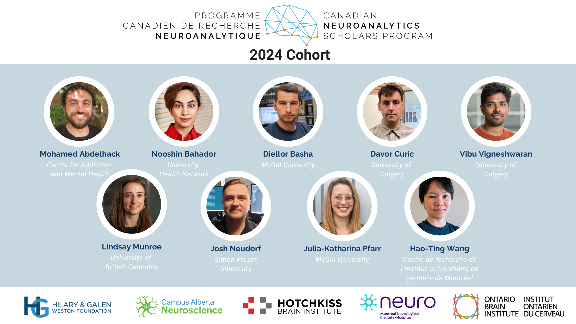
The Canadian Neuroanalytics Scholars Program (Programme Canadien de Recherche Neuroanalytique) is excited to announce its first cohort of Scholars from across Canada.

August 22, 2024

First Canadian Neuroanalytics Scholar Program Cohort Announced
The Canadian Neuroanalytics Scholars Program is made possible through partners including Campus Alberta Neuroscience, the Hotchkiss Brain Institute, the Tanenbaum Open Science Institute at The Neuro, and the Ontario Brain Institute, with support from The Hilary & Galen Weston Foundation.

Canadian Neuroanalytics Scholars Program

Launched in 2024, the Canadian Neuroanalytics Scholars (CNS) Program will support and train up to 20 postdoctoral scholars in advanced analytics, providing them with hands-on experience. The program will leverage and connect the infrastructure, resources, and expertise available through research and industry partners across Canada. The goal is to cultivate a world-class talent pool that can effectively utilize the existing open neuroscience data and meet the growing demand for neuroscience research in the fields of artificial intelligence and machine learning (AI/ML).

The CNS Program will consist of two cohorts of scholars on a two-year term, for a total of up to 20 scholars over four years. The Program leverages existing programs offered by Training Partners, to ensure scholars have formal affiliation and access to training, high-quality resources, infrastructure, and mentorship to grow their skillset. Research Partners provide further training and support to ensure the scholars have a fluent understanding of the datasets, methods used for collection and curation, and the clinical context.

Applications for the next CNS Program cohort will open in 2025.

For more information on the CNS Program, please visit the competition webpage.

Learn more about Cohort 2 of the CNS program here.

Cohort 1: Our 2024 CNS Scholars

The CNS Program is pleased to announce the extraordinary Scholars selected for the first cohort:
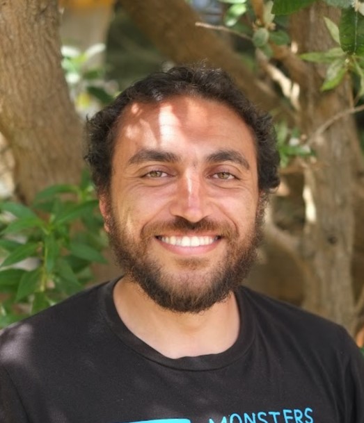
Mohamed Abdelhack, PhD – The Centre for Addiction and Mental Health (Toronto, ON)

Project title: Uncovering shared neural signatures of cognition in neurodegenerative disorders with neuroimaging data analytics and computational modelling

Neurodegenerative diseases affect a large percentage of elderly Canadians and manifest in various cognitive and motor symptoms depending on the affected brain area. Neurodegenerative diseases and vascular disorders may lead to types of dementia due to the effect of blood supply to the brain on neuronal health. Due to the interconnectedness of the brain, there are usually many shared symptoms that could result from similar impairments in brain processing circuits. However, we do not fully understand how cardiovascular illness may predispose individuals to dementia at the level of brain function. Previous studies found that Alzheimer disease, the most common neurodegenerative disease and cause of dementia, is associated with localized problems in areas related to memory and planning. In this study, I plan to use large datasets of brain imaging data and computational modeling to uncover shared and unique patterns of brain function between neurodegenerative illness and cardiovascular disease. I will build statistical models to find the relationships between cognitive performance and brain structural and functional measures. By understanding the statistics of how brain structure and function differs as cognitive changes develop, we can reveal which brain circuits are being affected across multiple disorders. This can enable us to build a mathematical model that we can validate using real patient data. This model will replicate the neural activation changes associated with various impairments and will enable us to understand how brain regions communicate with each other and how breakdowns in communication, due to a number of factors including cardiovascular disease, may lead to dementia. This will allow us to discover the early signs of neurodegeneration, allowing earlier intervention. It will also serve as a tool to monitor the effectiveness of therapies and their potential side effects.

Nooshin Bahador, PhD – University Health Network (Toronto, ON)

Project title: Developing biomarker for detecting seizure onset zone and monitoring seizure development
Epilepsy presents as recurrent seizures, characterized by abnormal and hyperactive neural firing in the brain. Monitoring these seizures often involves observing electrical activity through specific tests like EEG, a non-invasive method detecting surface brain activity, and LPF, an invasive technique assessing deeper brain structures. Seizures can vary in dynamics, evolving over time and potentially increasing in severity. Furthermore, seizures manifest diversely, originating from different brain locations and having various underlying causes. Accurate diagnosis of seizure type is crucial for effective treatment planning. Currently, there’s no objective measure to quantify changes in electrical brain activity during seizures, nor a standard measure aiding in identifying their root causes. Additionally, the mechanism of action varies among anti-seizure drugs. Predicting which medication will be effective in restoring normal brain electrical activity is challenging, and the degree of effectiveness from the perspective of brain electrical activity is currently not quantifiable. We propose that the characteristics of the chirp pattern (narrow-band frequency modulation pattern occurs during a seizure) can serve as a measurable indicator. Distinctive nature of chirp pattern makes it both unique and detectable that can stand out from background activity. So, the primary objective of this research is to develop an analytics tool that can automatically and reliably identify and characterize various types of chirp patterns in both animal models and human subjects. As we measure this pattern, we may not only observe changes but also discern how these changes relate to the mechanism of action of the anti-seizure drugs. The unique characteristics of chirp may also be useful in identifying false positive drug candidates that appear effective in animal trials but fail to work in humans.

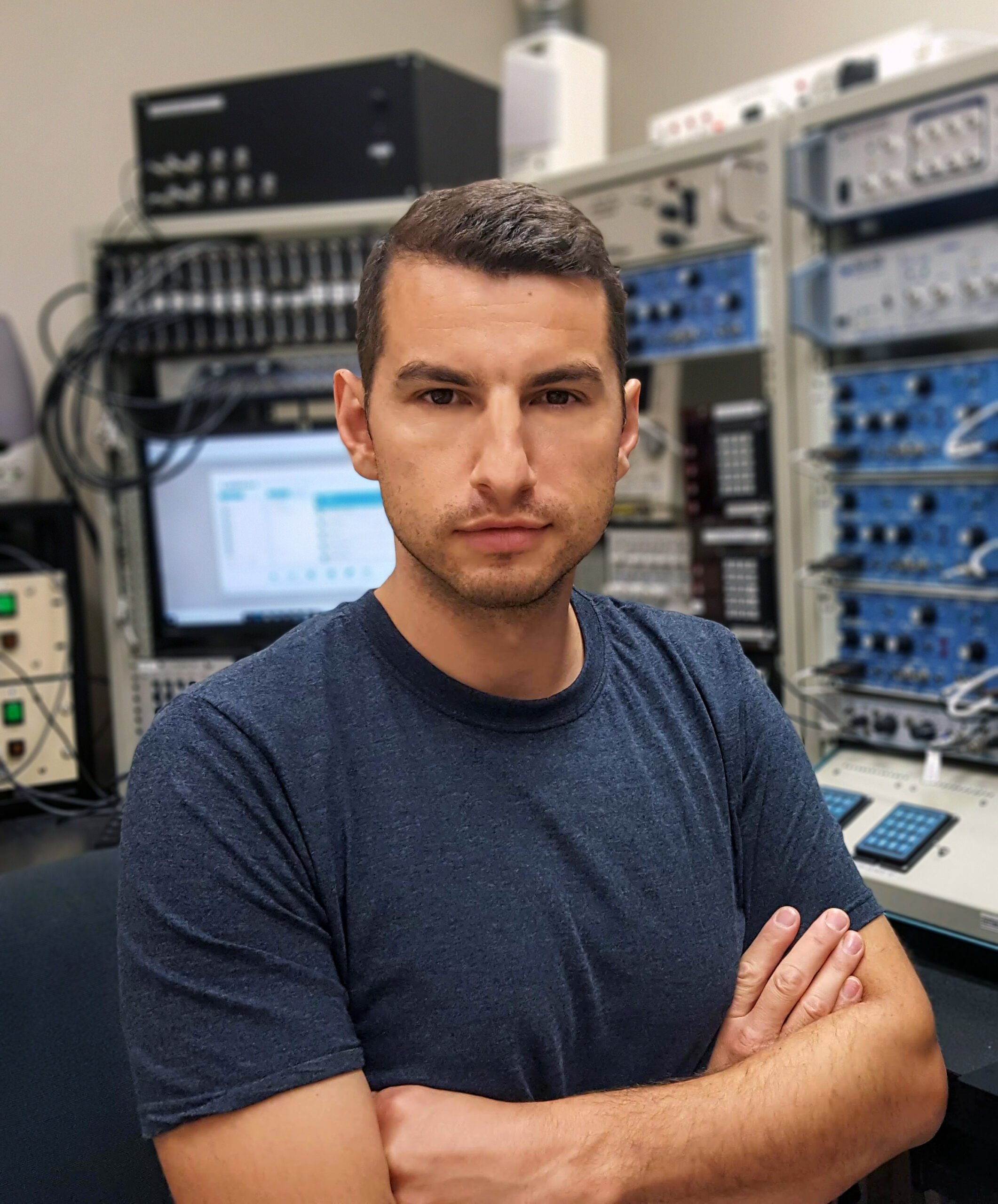
Diellor Basha, PhD – McGill University (Montréal, QC)

Project title: Leveraging Open, Multimodal Data to Forecast Risk and Resilience in Alzheimer’s Disease
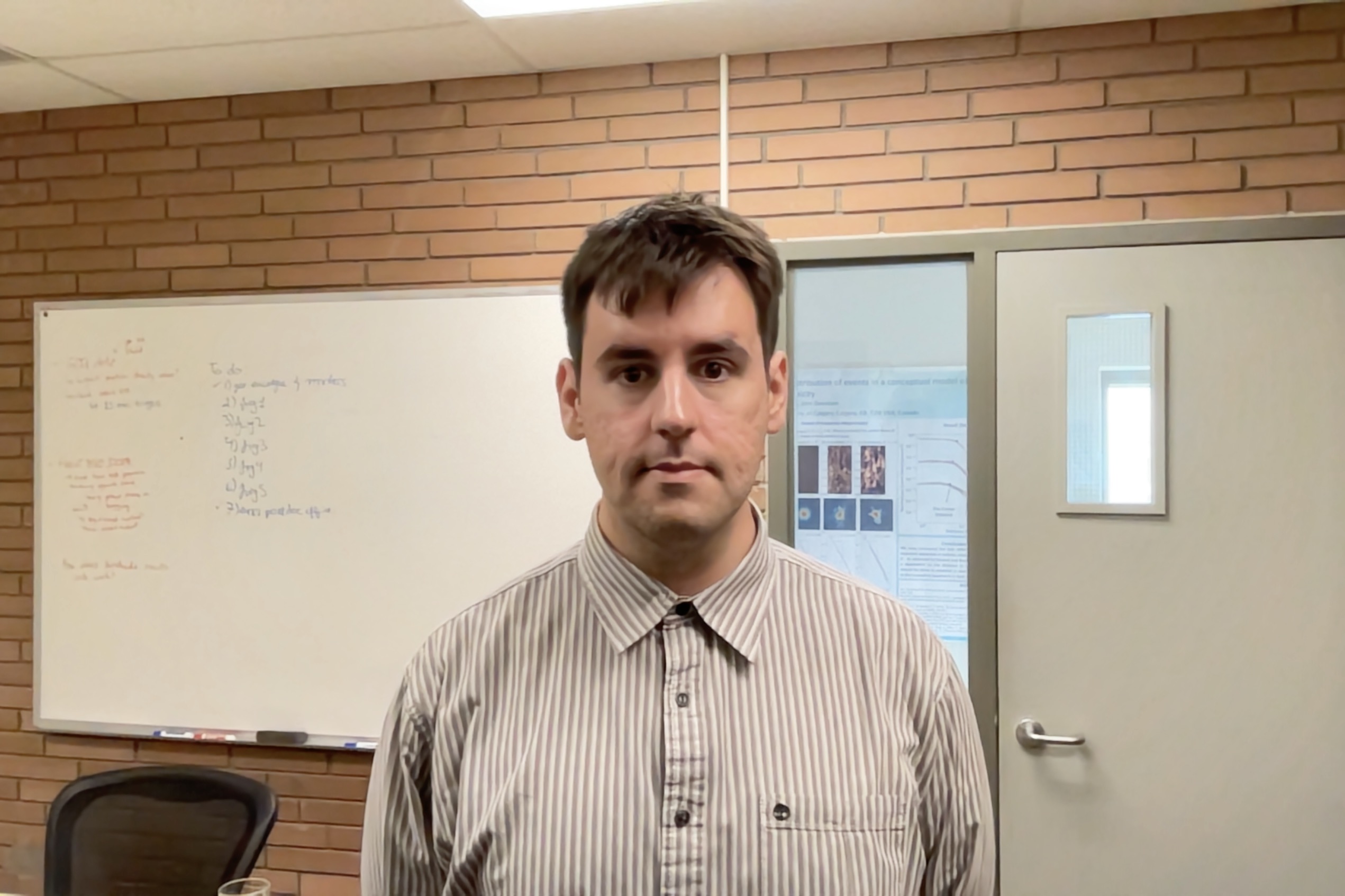
Alzheimer’s disease is the most common cause of dementia, accounting for 60%-80% of cases in Canada and worldwide. AD is characterized by cognitive decline, evidenced by memory loss, impaired reasoning and mood changes. With the aging of the population, AD cases are expected to triple by 2050. AD is marked by the buildup of harmful proteins, beta-amyloid (Abeta) and tau, in the brain, leading to the death of brain cells and widespread disruption of brain functioning. Efforts in early detection, like finding neurophysiological biomarkers of AD, are a top research focus. Recent years have seen an unprecedented surge in the availability of open neuroscience data, thanks to government and private initiatives. PREVENT-AD is such an initiative supported by the Canadian Open Neuroscience Platform, which has collected extensive brain imaging data and rich medical information over several years from healthy participants with family history of AD at risk of developing the disease. PREVENT-AD includes magnetoencephalography (MEG), electroencephalography (EEG), positron emission tomography (PET), magnetic resonance imaging (MRI), cognitive evaluations, genetic profiling, and blood samples. Harnessing the full research potential of these complex datasets requires advanced data science techniques designed for large-scale studies and made freely available to the research community. The primary aim of my project is to develop a practical open-source software toolkit specifically designed for staging, understanding, and treating AD using data from open repositories such as PREVENT-AD. The proposed Brainstrorm AD Module will allow researchers to interact with open data repositories and provide an environment for integrating the analysis of MEG, EEG, PET and MRI data. The AD Toolkit will be developed as a module within Brainstorm, a widely used open-source software for the analysis of neurophysiological and structural MRI data (>43,000 registered accounts and >3,500 publications). Current developments of the app include the integration of more imaging modalities, including PET – a major imaging tool used in AD clinics and research. I will leverage the PREVENT-AD data to identify electrophysiological markers that forecast AD risk and resilience in healthy subjects. The data available enable using artificial intelligence techniques, adapted from natural language processing applications, to capture subtle changes in brain recordings that signal the early build-up of tau and Abeta, before participants start presenting symptoms. The unique longitudinal data of PREVENT-AD will then be used to train a deep learning model to capture anomalies in whole-brain pattern of electrophysiological activity and forecast the risk of upcoming cognitive decline. Through this project, I will deliver a robust, user-friendly software tool that will be available to all researchers worldwide to validate and replicate our research results and advance their own investigations of AD.

Davor Curic, PhD – University of Calgary (Calgary, AB)

Project title: A Tangent Space Analysis for Early Detection and Progression Tracking of Neurodegenerative Disease

A major impediment to our ability to detect individuals who will progress to developing a neurodegerative disorder, is that pathological signatures (e.g., Tau, B-amyloid, alpha-synuclein) can manifest in the brain decades before symptom onset. As such, It is critical that we develop means of identifying changes to neural function before symptom onset, and that this method be non-invasive and scalable. One approach is to use the functional connectome (FC), a matrix representation of brain activity which can be obtained from neuroimaging modalities such as functional MRI scans in humans, and other modalities of sampled neural activity in model species, such as calcium imaging. The FC captures the interactions between brain regions, and changes could serve as biomarkers for early detection, disease progression, and treatment outcomes. The study of disease progression requires longitudinal data and a notion of a distance between FCs. This latter point is difficult as FC space is curved, not flat. Just as one needs to account for the Earth’s curvature to find the distance between two cities, the curvature of FC space needs to be considered to measure FC distances. Traditional metrics like correlation distances ignore this curvature, resulting in erroneous distances. ‘Geometry-aware’ approaches, like Tangent Space Analysis (TSA), have recently emerged that do take into account the curvature of the space. My research will utilize TSA to study the progression of Alzheimer’s disease (AD). Initially, I will use data from a mouse model engineered with mutations associated with the development of AD (5xFAD) to determine whether TSA can detect pathological changes related to AD, earlier and more accurately than traditional methods, and identify which brain areas are most affected. This data has been recorded using calcium imaging of the neocortical surface by the group of Dr. Alexander McGirr. The model species will be provide a controlled environment for testing TSA while human fMRI data is being processed and quality checked. Next I will extend TSA to human patients using open databases. For example, the Ontario Neurodegenerative Disease Research Initiative, PResymptomatic EValuation of Experiment or Novel Treatments (PREVENT) for AD, and the Canadian Consortium on Neurodegeneration in Aging datasets feature longitudinal fMRI data from individuals at risk of neurodegenerative disease. This would allow me to contrast individuals who develop dementia against those that do not, and then to track FC changes in relation to disease progression. If successful, this project could provide a non-invasive approach to studying neurodegenerative disease progression using novel data to develop methods to then deploy on existing open science datasets within the term of the CNS program. This would not only provide researchers with a new tool for studying the disease but also offer healthcare providers a new diagnostic tool to enhance patient care.

Lindsay Munroe, PhD – University of British Columbia (Vancouver, BC)

Project title: Predicting progression of neurodegenerative diseases with explainable AI

Neurodegenerative diseases such as Alzheimer’s disease (AD) and Multiple Sclerosis (MS) can have a profound impact on quality of life, yet predicting how these conditions will progress remains a significant challenge. Accurate prognosis is becoming increasingly important as new treatments emerge and as clinicians seek to deliver more personalized care. This project is developing advanced artificial intelligence (AI) tools to predict disease progression in individuals with AD and MS using longitudinal MRI scans and other health data. Unlike many AI systems that function as “black boxes,” this research uses explainable AI, an approach that not only generates predictions but also provides insight into how those predictions are made. The project will train and evaluate state-of-the-art AI models using large-scale datasets from Alzheimer’s and MS research studies. By combining brain imaging with clinical, genetic, and blood-based information, the models aim to improve the accuracy of disease forecasting while identifying the factors most closely associated with progression. In addition to predicting future clinical outcomes, the AI system will generate visual disease maps that highlight brain regions and features linked to disease changes over time. These insights could help uncover new biomarkers of progression, support earlier and more informed treatment decisions, and improve patient selection for clinical trials. As one of the first efforts to apply explainable AI to the prediction of MS progression from longitudinal neuroimaging data, this research has the potential to advance both the understanding and management of neurodegenerative disease.

Josh Neudorf, PhD – Simon Fraser University (Vancouver, BC)

Project title: Using Graph Neural Network Deep Learning to identify brain connectivity biomarkers for mild cognitive impairment and dementia in the aging population

The global population is aging rapidly. We are at a crucial juncture for understanding and addressing cognitive decline in this population. Dr. Randy McIntosh and I have identified changes in the structural and functional networks of aging brains associated with cognitive changes in healthy aging. I will begin a novel project with Dr. McIntosh to investigate how the rewiring of brain connections may lead to functional changes and act as biomarkers for mild cognitive impairment and dementia. These biomarkers will be identified using an artificial intelligence (AI) approach called Graph Neural Network (GNN) Deep Learning, which combines deep learning and graph theory (math for networks), to predict stages of mild cognitive impairment and dementia from brain network configurations. GNN Deep Learning maintains a network representation of the data as a graph rather than forcing the data to fit an inappropriate model, and the parameters needed for accurate prediction can be kept much smaller in the process to improve generalizability and training time. This model can integrate structural and functional connectivity in the same model, utilize brain region attributes (e.g., graph theory degree measures, neurotransmitter expression data from openly available neuromaps data, cortical thickness, fMRI activation intensity, etc.), and consider multiple individual-level variables simultaneously (e.g., age, sex, gender, socioeconomic status, ethnicity, physical activity, sleep quality, etc.) to create a comprehensive and diversely applicable model of the relationship between brain networks and the progression of dementia. There are advanced methods designed for GNN Deep Learning that can probe the model to identify specific elements in the network that drive the prediction (e.g., GNNExplainer), which would provide important insights for health professionals. I have experience applying GNN Deep Learning models to theoretical questions about how structural connectivity constrains functional connectivity, in addition to experience applying graph theory and dynamic functional connectivity approaches to studying preserved vs. declining cognitive ability in healthy aging. My experience has prepared me for this novel project at the intersection of AI and neurodegeneration research, identifying biomarkers for the progression towards mild cognitive impairment and dementia. These methods will be applied to open datasets from the Ontario Neurodegenerative Disease Research Initiative (ONDRI), Alzheimer’s Disease Neuroimaging Initiative (ADNI), UK Biobank, Human Connectome Project Lifespan, and Cambridge Centre for Ageing and Neuroscience (Cam-CAN). These brain models will be tailored to age, sex, gender, ethnicity, and socioeconomic status, and shared with health professionals to aid them in identifying risk factors for dementia, allowing for early interventions to protect and maintain healthy brain function.

Julia-Katharina Pfarr, PhD – McGill University (Montréal, QC)

Project title: Identifying Brain Targets for Major Depressive Disorder Research

Mental health conditions like depression have a tremendously high burden on an individual, the care-givers, the society, and economy. Mental disorders are common, they often onset early in life and they can persist across the entire lifespan. As of today, depression by itself causes 10% of all disability worldwide. Additionally, depression is highly heterogeneous: two people with depression can have an entirely different set of symptoms and very different underlying biological or environmental causes. We therefore need reliable and quantifiable disease markers. As depression is thought to be a brain disorder, brain imaging techniques hold promise in the search for depression disease markers. However, research in this field shows fundamental limitations which resulted in the fact that to this day no reliable biomarkers could be found. One limitation lies in the study design of previous studies which is known as a ‘case-control’ framework. This framework makes the assumption that the group of depression patients is a well defined group and distinct from a control group. This, however, is not a valid assumption and thus introduces a tremendous statistical bias already at the study design stage: which effects in the brain data are actually important for the disorder cannot be detected by fitting people into broad categories. One way to overcome this limitation is to let the brain data “sort itself out” or “learn its structure on itself” without making a priori assumptions about the data’sstructure, and find the relation with depression subtypes defined by the clinical variables. Identifying the most important brain features and modeling their relation to crucial phenomenological and clinical variables enables us to move away from making statements about groups toward making statements about individuals, i.e., precision psychiatry. With this project we attempt to find the most important brain features for depression biotypes, i.e. brain targets for depression. We aim to do so by harmonizing multimodal clinical and phenotypic as well as multimodal brain imaging data from different large-scale datasets. We will apply ensemble clustering on clinical and phenotypic variables to find comprehensively described depression subtypes to then use those as labels for self-supervised learning and find robust and reliable brain targets. We hope that with the combination of advanced computational methods and extensive external validation, we can identify brain targets meaningful for depression which could be reused in future studies to model norms and deviations for individuals with depression.

Vibu Vigneshwaran, PhD – University of Calgary (Calgary, AB)

Project title: Causal Deep Learning for Neuroimaging applications

The current exponential increase in data acquisition in all aspects of our lives, coupled with the continuous advancement in computational power, has led to many exciting innovations powered by machine learning (ML). Notably, the artificial intelligence field has witnessed a quantum leap with the emergence of deep learning, which has shown remarkable abilities in many important tasks, often surpassing traditional ML approaches and even human performance. In healthcare settings, brain imaging is one of the largest data contributors, which holds significant importance in the prevention, diagnosis, and treatment planning of neurological and mental diseases. Deep learning models have the potential to revolutionize and support neuroimage analysis by automating the identification of highly complex patterns in neuroimaging data. However, one of the challenges in adopting these tools in clinical radiology practice is that many existing models only identify and use correlations in the data without considering the underlying causal relationships. Unfortunately, models using such correlations often identify and encode biases present in imaging data that are not clinically meaningful, such as differences related to the population or technical image acquisition. This can lead to deep learning models learning shortcuts and biases, making it challenging for them to provide meaningful explanations for their decisions and generalize to new unseen data. Thus, the primary objective of this work is to develop and evaluate a flexible and novel framework that combines causality aspects and deep learning to not only deliver precise predictions and classifications but also provide valuable insights about the causal connections between clinical data and neuroimages. This framework specifically aims to identify causal relationships between non-imaging factors (such as disease state, genetics, lifestyle factors, and acquisition parameters) and complex neuroimaging datasets. The second objective is to extend this framework with the ability to generate realistic images for alternate realities. For example, using this ability, we could simulate what a brain image would look like if the patient did not have dementia and could visualize the differences. Furthermore, this framework would enable the identification of causal factors related to specific neurological and mental diseases and investigate how clinical variables change the appearance of the brain, which can be used for in-silico disease modelling and obtaining deeper insights about causal disease mechanisms. In summary, this project will develop a flexible framework for the automatic analysis of brain images and clinical variables that can provide direct explanations and an effective means of bias mitigation, deeper insights into disease mechanisms, and potential avenues for intervention across a range of neurological and mental diseases

Hao-Ting Wang, PhD – Centre de recherche, Institut universitaire de gériatrie de Montréal (Montréal, QC)

Project title: Predicting presymptomatic features of Alzheimer’s disease with transfer learning in the PREVENT-AD cohort

Recent research suggests significant overlap in the brain changes underlying various neurodegenerative conditions, including Alzheimer’s disease, schizophrenia, and Autism Spectrum disorder. Traditionally these conditions are studied in isolation, but we believe these conditions can be better investigated together as a broad spectrum. This project explores a novel approach to investigate these commonalities by pooling multiple datasets together. Once pooled, we aim to showcase how using a model pretrained on this big datasets and a machine learning technique called transfer learning, we can predict diagnoses within a smaller clinical dataset, namely in the PREVENT-AD dataset. Ultimately, the goal is to discover markers that can predict the development of cognitive decline. This analysis will focus on identifying “transdiagnostic brain signatures”: shared features across these conditions. Brain scans will be used to extract relevant information. A pre-trained model, informed by data encompassing natural variations in brain health across a large population, will then be applied to a dataset specifically focused on Alzheimer’s development. The model architecture chosen leverages knowledge of brain connectivity to understand how these features evolve over time. Importantly, it also produces interpretable results, allowing researchers to dissect the model’s findings. Furthermore, a secondary aim is to develop a user-friendly software framework to help streamline future research by enabling seamless analysis of brain scans from diverse datasets. This bridge between research and clinical application has the potential to improve our understanding and management of neurodegenerative conditions. The research will be conducted at the SIMEXP lab at the CRIUGM under the supervision of Professor Pierre Bellec, a brain imaging and deep learning expert, and in collaboration with Dr. Sylvia Villeneuve, an expert on Alzheimer’s disease and the PREVENT-AD dataset at McGill University. My experience in software development is supported by my recent contribution to the Python library NiLearn and deep learning experience supported by previous postdoc projects.

Generously funded by:

The goal of The Hilary & Galen Weston Foundation is to contribute to charities whose bold ideas shape a better future for everyone. For more information, please visit hgwf.org

This program is made possible through the support of our partners:

Campus Alberta Neuroscience (CAN), established in 2012 with support from the Government of Alberta, is a province-wide neuroscience network connecting the Universities of Alberta, Calgary and Lethbridge to increase the impact of neuroscience and mental health research, education and translation, developing the province as an epicenter for neuroscience excellence. For more information, please visit albertaneuro.ca.

The HBI’s vision is “Healthy brains for better lives”. Their mission is to inspire discovery and apply knowledge towards innovative solutions for neurological and mental health disorders. This mission is guided by six core values: Excellence, collaboration, integrity, impact, creativity, and relevance. For more information, please visit hbi.ucalgary.ca

The Neuro (Montreal Neurological Institute-Hospital) is a bilingual academic healthcare institution. We are a McGill research and teaching institute; delivering high-quality patient care, as part of the Neuroscience Mission of the McGill University Health Centre. They  are proud to be a Killam Institution, supported by the Killam Trusts. For more information, please visit mcgill.ca/neuro

The Ontario Brain Institute (OBI)  is a provincially funded, not-for-profit organization that accelerates discovery and innovation, benefiting both patients and the economy. OBI works to establish Ontario as a world leader in brain research, commercialization and care. For more information, please visit braininstitute.ca

The CNS program is pleased to collaborate with the following Canadian centres of excellence in AI/ML as training partners: