April 15, 2026

Second Canadian Neuroanalytics Scholar Program Cohort Announced
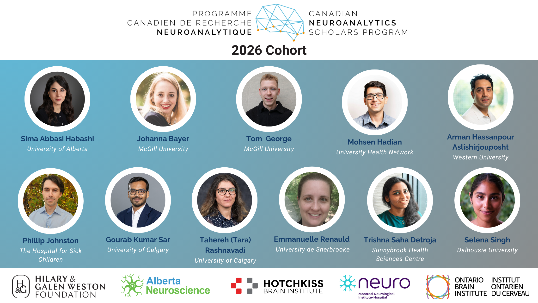
The Canadian Neuroanalytics Scholars Program is made possible through partners including Alberta Neuroscience, the Hotchkiss Brain Institute, The Neuro, and the Ontario Brain Institute, with support from The Hilary & Galen Weston Foundation.

Canadian Neuroanalytics Scholars Program

The Canadian Neuroanalytics Scholars (CNS) Program is proud to announce the selection of its second cohort of scholars, continuing its mission to strengthen Canada’s leadership in advanced data analytics and utilization of open neuroscience datasets.

Launched in 2024, the Canadian Neuroanalytics Scholars Program supports and trains postdoctoral scholars in advanced analytics, providing hands-on experience in cutting-edge computational methods. The program leverages and connects infrastructure, resources, and expertise across leading research and industry partners nationwide. Its goal is to cultivate a world-class talent pool capable of effectively utilizing open neuroscience data and meeting the growing demand for neuroscience research in artificial intelligence and machine learning.

This year, eleven scholars have been selected to join the program. The cohort will work across nine institutions spanning the country, from the East Coast to Alberta, reflecting the program’s national reach and collaborative model.

With scholars embedded within Canada’s leading neuroscience hubs, the program provides early-career postdocs with the unique opportunity to connect and work with research and industry partners across Canada while they gain interdisciplinary technical and translational skills.

The Canadian Neuroanalytics Scholars Program is made possible through the partnership of Alberta Neuroscience, the Hotchkiss Brain Institute, The Neuro (Montreal Neurological Institute-Hospital), and the Ontario Brain Institute, with generous support from The Hilary & Galen Weston Foundation.

Together, these partners are advancing a coordinated national training platform that strengthens Canada’s neuroscience ecosystem while accelerating innovation at the intersection of brain science and advanced analytics. By harnessing the power of open neuroscience data, the CNS Program promotes collaboration, transparency, and shared scientific progress across institutions and disciplines. This collective, data-driven approach enhances rigor and reproducibility in research, positioning Canada to lead the broader knowledge and innovation economy shaped by artificial intelligence and machine learning.

For more information about the Canadian Neuroanalytics Scholars Program or press inquiries, please contact:

Rachel Paris, Project Manager, Alberta Neuroscience – rachel.paris@ucalgary.ca

For more information on the CNS Program, please visit the competition webpage.

Learn more about Cohort 1 of the CNS Program here.

Cohort 2: Our 2026 CNS Scholars

The CNS Program is pleased to announce the extraordinary Scholars selected for the second cohort:

Sima Abbasi Habashi – University of Alberta (Edmonton, AB)

Project title: Multi-Modal Machine Learning to Uncover Genetic and Immune Pathways Driving White Matter Hyperintensities and Cognitive Decline

As we age, many people develop white matter hyperintensities (WMH), which are small spots seen on brain scans that reflect damage to tiny blood vessels in the brain. These WMH are very common and often worsen over time, but some people are affected more than others. WMH are known to raise the risk of memory loss and dementia. Our goal is to understand why some individuals develop more WMH than others and how this contributes to cognitive decline. Previous research from our team has shown that people with fast-progressing WMH have different immune activity in their blood. We’ve also shown in laboratory models that immune cells can stick to small brain vessels and block blood flow, leading to further damage. These findings suggest that inflammation and blood vessel dysfunction may be key drivers of WMH and memory problems. However, we still don’t fully understand which genes or biological pathways are involved, or how to predict who is most at risk. In this project, we will use data from the Ontario Neurodegenerative Disease Research Initiative (ONDRI), a study of people with early signs of brain and memory disorders. This dataset includes brain scans, genetic information, and yearly memory tests. First, we will measure the size of WMH in each participant’s brain using advanced image analysis techniques. Then, we will scan their genetic data to find common genetic variants, called SNPs, that are linked to WMH severity and worsening over time. To make sense of the results, we will group these genetic variants into biological pathways, with a focus on inflammation and blood vessel health. This helps us go beyond individual genes to see the bigger picture of how complex systems in the body affect brain health. Next, we will use machine learning, a form of artificial intelligence, to combine genetic data, brain scan results, and memory scores. This will allow us to build models that can identify which combinations of factors are most useful for predicting who is likely to experience WMH progression and cognitive decline. We will also test newer approaches such as graph-based models that can detect interactions between genes and highlight key pathways involved in disease. Importantly, we will check whether certain risk patterns differ by age or sex. By the end of the project, we aim to develop a prototype risk score that can help identify people at higher risk of cognitive decline caused by inflammation and small vessel disease. This tool could eventually help doctors detect problems earlier, tailor treatment strategies, and guide clinical trials for new therapies. Our research may also reveal biological pathways that could be targeted by drugs to protect brain health and prevent dementia. This project uses cutting-edge data science to solve a critical challenge in brain aging and aligns strongly with the CNS Program’s mission to apply advanced analytics to real-world problems in neuroscience.

Johanna Bayer – McGill University (Montréal, QC)

Project title: Personalized deep longitudinal normative modeling for tracking Parkinson’s disease progression

Parkinson’s disease (PD) affects a substantial proportion of the population, manifesting in progressive motor and cognitive symptoms. Despite advances in neuroimaging, predicting individual disease progression remains a critical unmet need due to three challenges: 1) a complex, non-linear disease progression that makes predictions of individualized trajectories difficult, 2) the need to distinguish pathological changes from normal age-related brain variation, and 3) systematic biases introduced when pooling multi-site datasets which confound predictive models, especially for a disease that evolves over time. Methodology We will, for the first time, implement and combine two deep learning-based approaches to address these challenges. Longitudinal Velocity Modeling: By tracking individuals over time, we will construct personalized brain change trajectories from anatomical scans and derived measures and compare these against normative aging patterns to identify disease-specific progression markers that align with cognitive and motor decline in PD. Deep Normative Modeling: Using whole‐brain imaging data from multiple scans from >20,000 healthy individuals, we will identify latent patterns to establish comprehensive normative baselines. These capture subtle multivariate relationships beyond conventional univariate methods. We will contrast these patterns with data from over 2,000 individuals in the largest multi‐center PD cohort to link deviations to disease progression. Deep normative velocity modelling: For the first time, we will combine those two methods, which will allow us to create an unprecedented multimodal model of the normal variation of the structural neuroimage in healthy controls. This information will then be used to contrast brain changes in individuals with PD, further allowing to model disease progression in PD patients based on longitudinal changes. Dissemination of open source tools: We will disseminate all models and tools that were created as part of this research, including solutions to longitudinal modelling in multi-site data sets, in order to accelerate longitudinal research in neurodegenerative diseases. Significance and Impact This research addresses a critical gap in precision medicine for PD by applying advanced deep learning and longitudinal models to large, open neuroimaging datasets. The project will result in an age‐dependent representation of typical and atypical structural brain variation across multiple imaging modalities, including forward predictions of normal brain development for a period of choice. Further, by enabling us to quantify the significance of patient-specific disease progression in relation to symptoms of PD, our approach will help to optimize disease prevention and therapeutic interventions. The open-source dissemination of our methodological advances will establish new standards for longitudinal multi-site neuroimaging analysis and accelerate discovery across the field.

Tom George – McGill University (Montréal, QC)

Project title: Foundation Models for Neuroscience: from open brain data to neurogenerative disease prediction

Artificial intelligence has recently taken a major step forward. Tools like ChatGPT have captured the world’s imagination by showing a remarkably powerful ability to understand language. These models are trained with simple objectives on vast datasets, allowing them to learn the complex structure within. A crucial feature of this approach is that by learning the data’s underlying structure so deeply, the models can then perform well on new tasks which they were never explicitly trained for, making them true “foundation models”. Our project asks a straightforward question: What if we could use this same powerful approach to understand the human brain? Many of these models learn by exploiting “invariances” to uncover the essential meaning, or latent structure, within data while ignoring surface-level details. For example, a picture of a cat is still a cat even if some parts are hidden; its identity is an invariant property. When tasked with predicting the hidden parts of the image, a model must move beyond raw pixels and develop an abstract, internal representation—a core concept of a cat—forcing the model to capture the data’s fundamental structure, not just its superficial form. This makes its representation powerful and effective for subsequent downstream tasks which, in the case of a neural foundation model, may include predicting disease or classifying neural activity. Our project is based on the idea that brain activity has similar properties. The neural pattern for a memory, for example, should have a consistent structure each time it is recalled. We plan to build the first large-scale neuro-foundation model by training it on massive, open-source datasets of brain recordings. A key scientific goal is to discover which types of these neural invariances provide the most powerful signals for learning. The model will be tasked with various objectives—such as predicting activity in one brain region based on another, or across different moments in time—and we will systematically test which of these learning rules is most effective. This process will enable the model to learn the fundamental principles of brain activity without needing specific labels, unlocking the potential of vast, unlabeled datasets. Just as LLMs demonstrated for natural language, we believe the outcomes of this project could help usher in a new epoch for neuroscience research. We will produce a general-purpose model of brain activity applicable to numerous research and clinical problems. For neurodegenerative diseases like Alzheimer’s, the applications are direct. The model could be used to detect the earliest signs of disease in brain scans, long before symptoms appear. It could help predict how a patient’s condition might progress or propose best treatment options. By creating this foundational tool and making it open- source, this project aims to revolutionise translational neuroscience and accelerate the development of effective treatments for brain disorders.

Mohsen Hadian – University Health Network (Toronto, ON)

Project title: Prognosticating Alzheimer’s Disease Progression Using Multimodal Biomarkers: An Explainable AI Approach with ADNI

Alzheimer’s disease (AD) is the most common cause of dementia, affecting millions of people worldwide and placing immense emotional and economic burden on families and healthcare systems. The disease unfolds slowly, often beginning with subtle memory problems in a phase known as mild cognitive impairment (MCI). However, not everyone with MCI will progress to Alzheimer’s dementia. Identifying who is most at risk — and why — remains a major challenge in clinical care and research. This uncertainty limits our ability to provide timely interventions and slows the development of effective treatments. In this project, we aim to build an artificial intelligence (AI)–based model that can predict how Alzheimer’s disease will progress in individual patients. What sets this work apart is our focus on multimodal biomarkers — that is, biological clues collected in different ways. These include brain scans (like MRI and PET), genetic information (such as APOE4 status), fluid-based biomarkers (from spinal fluid and blood that includes tau, amyloid-beta, and neurofilament light), and clinical assessments of cognition and daily functioning. We will use data from the Alzheimer’s Disease Neuroimaging Initiative (ADNI), a globally recognized open-access dataset that tracks people across the full spectrum of cognitive aging — from healthy aging to MCI to dementia — with long-term follow-up. Our model will not only predict who is likely to decline, but also explain which combinations of biomarkers are most important for that prediction. This is done using a form of explainable AI, which helps translate complex algorithms into understandable and trustworthy insights. This work is especially timely because recent studies suggest that blood-based biomarkers may offer a simpler, less invasive way to detect Alzheimer’s-related changes. By comparing the value of different types of biomarkers — for example, blood versus spinal fluid versus imaging — our project could help determine which tests are most practical and informative in real-world settings. We will also explore whether there are different “subtypes” of disease, such as patients who decline due to metabolic or vascular changes, to improve personalized care. Ultimately, this project supports the CNS program’s goals by applying advanced analytics to open neuroscience data in a way that is transparent, reproducible, and clinically meaningful. Our findings could help future trials target high-risk individuals more effectively and offer new tools for early intervention and care planning. All code and findings will be shared openly to maximize impact. The project will also benefit from collaboration with the Toronto Dementia Research Alliance (TDRA), helping ensure that findings are translatable across real-world clinical settings in Ontario.

Arman Hassanpour Aslishirjouposht – Western University (London, ON)

Project title: Multimodal Machine Learning for Monitoring and Predicting Speech and Motor Symptoms in Neurodegenerative Diseases using Open Neuroscience Data

Neurodegenerative diseases, including Alzheimer disease and Parkinson disease, are progressive brain disorders that affect millions of individuals in Canada and worldwide. These conditions impair speech, movement, memory, and thinking abilities, often beginning with subtle changes that may go unnoticed for years. Because early symptoms can be challenging to detect using traditional tools, many individuals experience delays in diagnosis and intervention. Determining the disease course (rapid versus slow progression) also remains a clinical challenge, creating ambiguity for families. These delays can reduce treatment options and negatively affect outcomes. This project aims to create an intelligent and accessible monitoring system that can identify and track these symptoms earlier by combining multiple types of data, such as speech, movement, cognitive function, and neuroimaging, into a unified model. By analyzing how people talk, walk, and think, the system will capture early, often overlooked changes in behaviour. When linked to brain imaging, these signals provide a clearer picture of how neurodegenerative diseases develop and progress. The research will use three large, open neuroscience datasets: the Ontario Neurodegenerative Disease Research Initiative (ONDRI), the COMPASS-ND cohort, and the Brain-Eye-Amyloid Mobility (BEAM) study. These datasets contain thousands of assessments across diverse populations and disease stages. The project will utilize advanced artificial intelligence (AI) to identify key features that distinguish between conditions and predict individuals likely to progress more rapidly. The models will be optimized for transparency using explainable AI techniques, helping clinicians and patients understand how and why specific patterns are flagged. Outputs will be verified through checkbacks with healthcare professionals and individuals with lived experience to ensure they are clinically relevant. The system will be developed in collaboration with clinicians, data scientists, advocacy groups (including the Alzheimer Society, Parkinson Canada, and Heart & Stroke Foundation), and community members. Tools will be designed with adaptability and inclusivity in mind, enabling deployment in primary care, long-term care, and remote platforms. A key innovation of this work is its focus on an equitable, community-informed model design. Dataset bias across sex, race, and ethnicity will be addressed during training and evaluation. Voluntary contributions from underrepresented communities will help expand the model’s fairness and reach. The resulting tools will align with FAIR and Indigenous data principles where appropriate and will be open-source, transparent, and easy to integrate. This research will enable earlier intervention, informed planning, and more personalized care. It supports the CNS Program’s mission by advancing computational neuroscience and translating open data into impactful, scalable digital health tools.

Phillip Johnston – The Hospital for Sick Children (Toronto, ON)

Project title: Detecting Invisible Brain Injury from Repeated Head Impacts in Youth with Generative Modelling and Machine Learning

It is now well understood that mild traumatic brain injuries, also known as concussions, can have long term damaging effects on brain health. But many head impacts, such as those experienced regularly by contact sports players, deliver comparable force to the brain without producing telltale concussion symptoms, and therefore go largely undetected. Both types of injury have now been linked to cognitive impairment and, most concerningly, neurodegeneration. This includes progressive, untreatable brain diseases such as Chronic Traumatic Encephalopathy (CTE). Given the high incidence of head impacts in Canada and around the world, particularly among youth, there is therefore an urgent need to clarify how these asymptomatic (“subclinical”) head impacts damage neural circuits, and to detect this type of neural damage early to mitigate risk of permanent, progressive brain injury. With this project we aim to address this need by applying advanced neuroanalytic techniques to open brain signal data from a group of adolescent football players, and comparing them to a large open database of healthy controls. Brain signals were recorded with magnetoencephalography (MEG), a measurement technique which is highly sensitive to alterations in the activity of large groups of neurons, capable of detecting impact-related abnormalities before they show up as structural differences on typical clinical brain scans. However, while MEG provides rich information about ongoing neural activity, it cannot directly reveal the underlying disease processes. To address this we will use generative neurophysiological models – mathematical simulations of neural activity that can link observed MEG signals to otherwise unobservable physiological changes, like disrupted neurotransmitter levels. We will then use supervised machine learning to extract clinically-useful insights from the rich data that results, which will include conventional MEG measures and modelled physiological parameters estimated at many points across the brain. Specifically, we will estimate an individual’s degree of head impact using these brain data as predictors, using real measures of head impact from helmet-mounted sensors to train the model and verify (cross-validate) the results. In this way, we aim to establish an approach for young people who are concerned about neural damage, but who have not experienced a diagnosed concussion, to quantify their impact exposure from a single MEG recording. Doing so will deepen understanding of the basic mechanisms of neural dysfunction after subclinical head impact, and also create clinically-useful decision-support tools to mitigate the growing risks of neurodegeneration and long term cognitive impairment in young people.

Gourab Kumar Sar – University of Calgary (Calgary, AB)

Project title: Functional Role of Partial Synchronization in Neurological Diseases
It has been experimentally verified that synchronization and partial synchronization of brain activity play an important role in the pathogenesis of several neurological diseases, such as Parkinson’s disease, Alzheimer’s disease, and essential tremor (among others), as well as in biological and cognitive function. Yet, the precise mechanisms by which biological function and cognition emerge from the spatiotemporal patterns of (partially) synchronized neuronal activity remain largely unexplored. Initial work on the full human brain scale for healthy individuals has revealed that as different brain regions interact dynamically to perform cognitive tasks, coexisting synchronization and desynchronization among them – referred to as chimera states – can be observed. The ability to dynamically switch between different levels of integration (increased synchronization) and segregation (decreased synchronization) across different brain regions and, hence, different chimera states is believed to be crucial for cognitive performance and to be affected in neurodegenerative diseases: Excessive integration is often associated with conditions like epilepsy, while excessive segregation may impair cognitive flexibility, contributing to deficits seen in disorders such as schizophrenia or Alzheimer’s disease. The overall goal of this project is to investigate whether and how chimera states are affected by neurodegenerative diseases. Leveraging the expertise of the lab of Dr. Davidsen (supervisor) in computational neuroscience, network neuroscience, chimera states and neuroscience data and my extensive background in the mathematical theory and analysis of synchronization in biological systems, we will combine state-of-the-art mathematical models of neural interactions with high-resolution experimental open neuroscience datasets using a computational neuroscience approach centered around synchronization and chimera states. Our approach involves model simulations that leverage structural connectomes – matrices that represent patterns of connections between brain regions derived from neuroimaging techniques like diffusion MRI in humans. We will take advantage of open datasets such as PResymptomatic EValuation of Experiment or Novel Treatments (PREVENT) for Alzheimer’s Disease (AD) and those provided, for example, by the Alzheimer’s Disease Neuroimaging Initiative (ADNI), and the Canadian Consortium on Neurodegeneration in Aging (CCNA). Combining these datasets with our computational model approach will enable us to investigate the emergent synchronization patterns in silico in relation to disease progression. This will allow us to establish the possible connection of chimera states with neurological diseases and more broadly cognitive abilities. The latter will be achieved by leveraging data beyond structural connectomes such as functional connectomes and most importantly performance data for cognitive tasks, which are part of CCNA, for example, as well.

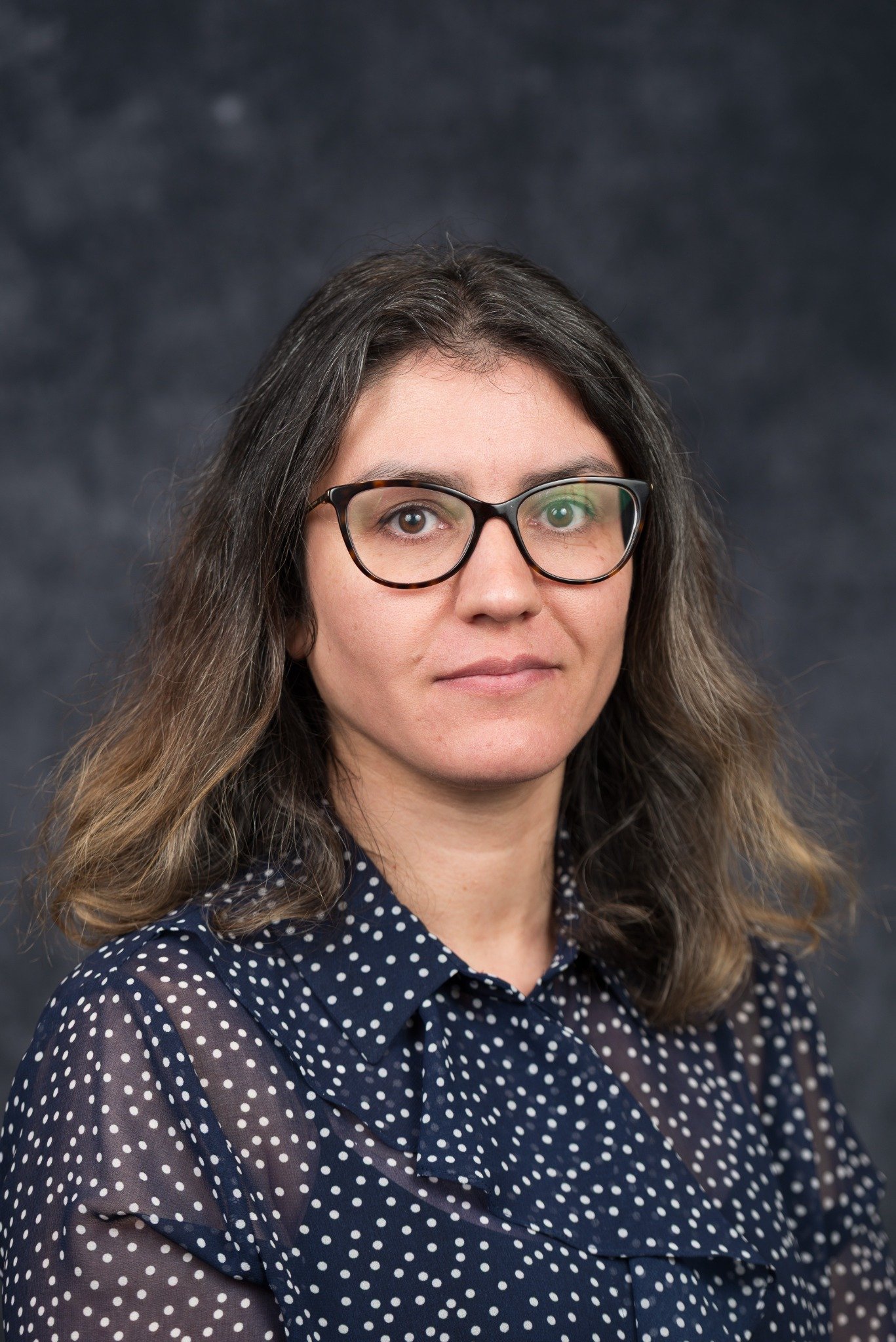
Tehereh (Tara) Rashnavadi – University of Calgary (Calgary, AB)

Project title: Causal Network Inference in Drug-Resistant Epilepsy Using Information-Theoretic Granger Causality with Neural Network-Based Feature Learning on Intracranial EEG-fMRI

Epilepsy is a serious brain disorder affecting over 50 million people worldwide. While many patients benefit from medication, a significant number—those with drug-resistant epilepsy—continue to experience seizures that greatly affect their quality of life. One of the biggest challenges in treating these patients is accurately identifying where seizures start in the brain and how they spread. Current methods are limited, especially because they can only record activity from a small number of brain regions using implanted electrodes. This project aims to overcome that challenge by developing a new AI-powered framework to map how different brain regions influence one another over time—essentially identifying “who talks to whom” in the brain during seizure activity. To do this, we will use an advanced technique called Granger causality, which can help determine if activity in one brain region predicts future activity in another. By combining this method with powerful machine learning tools like neural networks, we will be able to analyze complex brain recordings more accurately and at a much larger scale. We will apply these tools to a rare and valuable dataset: simultaneous recordings of electrical brain activity (intracranial EEG) and brain imaging (fMRI) from 70 patients with epilepsy. Using this combined data, we will create maps of how seizures propagate across the brain, even in regions not directly recorded by electrodes. These maps could significantly improve how we identify seizure onset zones and guide surgical decisions for epilepsy patients. Beyond epilepsy, we will also test whether this approach can help understand how brain networks change in aging and neurodegenerative diseases like Alzheimer’s. For this, we will use open-access brain imaging datasets such as the Alzheimer’s Disease Neuroimaging Initiative (ADNI). By comparing brain network patterns between healthy individuals and those with early signs of dementia, we hope to uncover changes in brain communication that could one day help with early diagnosis or monitoring of disease progression. Ultimately, this research will create a reusable, interpretable, and clinically relevant framework for mapping brain network dynamics. It could lead to better surgical planning in epilepsy and open new pathways for understanding and treating neurodegenerative conditions, aligning with the CNS Program’s mission to support innovative, data-driven neuroscience.

Emmanuelle Renauld– Université de Sherbrooke (Sherbrooke, QC)

Project title: Efficient classification of chronic back pain via minimal data aggregation from multi-site multi-modality open datasets

Chronic pain is a major health concern affecting nearly 8 million Canadians, and one of the most common types is chronic back pain (CBP). In 2021, the World Health Organization officially recognized chronic pain as a disease, highlighting the urgent need for better ways to understand and treat it. Research has shown that chronic back pain can lead to changes in the brain, but so far, these changes haven’t been consistent enough to reliably identify people with CBP using brain scans. Our project aims to change that by finding a small, reliable set of brain-based markers (called “biomarkers”) that could help diagnose chronic back pain. These biomarkers will be identified using different types of magnetic resonance imaging (MRI), a non-invasive way to look at the brain’s structure and activity. To do this, we will gather brain scan data from about 800 people with chronic back pain, using both open-access research databases (such as openpain.org, painrepository.org, and openneuro.org) and data collected by our lab and research collaborators. This will make our study the largest of its kind. For comparison, we will also use brain scans from people without chronic pain, carefully matched by age and sex, using open datasets from the Montreal Neurological Institute or the Human Connectome Project. The MRI techniques we will use include structural imaging (to look at brain anatomy), diffusion imaging (to examine brain wiring), and functional MRI (to measure brain activity at rest). We will use the most advanced and reliable methods currently available to process the brain scans and extract important information from them. Because the data comes from many sources and scanners, we will use advanced methods to standardize the information so it can be meaningfully compared. Next, we will use computer algorithms that can find patterns in data to build and test models that can tell whether someone has chronic back pain based on their brain scans. We will carefully train these models using some of the data, and then test their performance on new data to see how accurate and reliable they are. Our ultimate goal is to develop a diagnostic tool that can identify chronic back pain from brain scans with high accuracy. In the long run, this research could not only help doctors diagnose CBP more effectively, but also lead to better treatments including both medications and non-drug therapies, tailored to how chronic pain affects the brain. By improving how we understand and identify chronic back pain, this project has the potential to transform the lives of millions of Canadians and others worldwide who suffer from this often invisible and debilitating condition.

Trishna Saha Detroja – Sunnybrook Health Sciences Centre (Toronto, ON)

Project title: Unraveling the role of sleep-related cortical cell types subpopulations in sleep, dementia-related brain changes, and dementia in aging

Dementia, a syndrome marked by cognitive decline and inability to carry out activities of daily life is an emerging preeminent public health issue in Canada and worldwide, with a prediction to increase by 51% between 2020 to 2030. Since no effective treatment currently exists, advancing our understanding of dementia aetiology is utmost important to elucidate and target causal risk factors. Sleep is pivotal for maintaining brain health and cognition. Research has shown disrupted sleep is linked to increased risk of dementia and neurodegeneration, making it a potentially modifiable target for dementia prevention. However, sleep play as double-edged sword, where poor sleep cause dementia while dementia worsen sleep. Current understanding of biological process underlying this vicious cycle is limited, hindering therapeutic intervention to prevent adverse effect of poor sleep on cognitive decline and vice versa. Thus, my research aims to address this gap, identifying cellular and molecular changes in brain driving this bi-directional relationship between sleep and dementia. Brain contains eight major cell types: astrocytes, microglia, oligodendrocytes, oligodendrocyte precursor cells, excitatory neurons, inhibitory neurons, endothelial cells, and pericytes. These cell proportions shift with dementia and aging. I hypothesize that identifying changes in brain cell type proportion that link sleep fragmentation to brain structure changes and to cognitive decline will unveil underlying mechanisms. To bridge this knowledge gap, I have two specific goals. First, I’m aiming to identify sleep fragmentation-related cell type changes associated with macroscopic brain changes and dementia. Second, I will apply a Mendelian randomization approach to decipher causal direction between sleep and dementia. To achieve this, we utilized machine-learning deconvolution method to infer brain cell-type proportions from bulk RNA-Seq data and linked these proportions to wearable sensor-based measures of sleep fragmentation. We also conducted Genome Wide Association Studies on sleep fragmentation data from UK Biobank and brain-cell type proportions from the Rush Memory and Aging Project. Our preliminary data showed differential association between cell type proportion, sleep measures and dementia pathologies; for instance, some microglia cell types are negatively associated with sleep fragmentation while positively correlated to dementia related pathologies. Altogether, my research will deploy multi-modal datasets including wearable measurement of sleep fragmentation, brain single-nucleus and bulkRNA-Seq, genotype, plasma proteome and brain MRI data from multi-cohort studies to map causal link between sleep, cell composition changes, brain structure and dementia. This will determine whether sleep fragmentation causes neurodegeneration or results from it, guiding new therapeutic development to prevent dementia targeting either sleep fragmentation or its cellular consequences.

Selena Singh – Dalhousie University (Halifax, NS)

Project title: Predicting Treatment Response and Relapse in Major Depression Using EEG Signatures of Neural Attractor States

Major depressive disorder (MDD) is the leading cause of disability worldwide, affecting ~10% of adults. Depression recurrence (known as “relapse”) is common, with rates between 35–85%. Also, treatment resistance affects nearly half of patients. Clinicians need tools to predict who will respond to certain treatments, and when someone might relapse. The existing tools are, however, limited, and we still do not have a complete understanding of depression’s neurobiology. This project will investigate whether depressive states and relapse are driven by stable, “sticky” brain activity patterns that resist change, known as “attractor” states, and whether we can use these patterns to predict treatment response and relapse. Under the supervision of Dr. Abraham Nunes and in collaboration with the Canadian Biomarker Integration Network in Depression (CAN-BIND), I will investigate these questions using brain activity data from two datasets: 1) CAN-BIND-1, a completed study of antidepressant response in MDD; and 2) CAN-BIND-20, an ongoing study of relapse in patients who have recovered from MDD. Attractor states have two properties: 1) resistance to change even when challenged, and 2) activity tends to settle or “converge” into them over time. To study the first property, resistance to change, I will analyze data from the CAN-BIND-1 study, which included 211 people with MDD, and 112 healthy volunteers. Patients completed an 8-week course of antidepressants, and their brain activity was measured using electroencephalography (EEG; a non-invasive way to record brain activity) before and after treatment. Participants also completed tasks that tested attention, emotional reactions and learning from rewards. These tasks act as natural “challenges” for the brain. I will test whether patients whose brain activity patterns remain rigid during these tasks are less likely to respond to antidepressants. The second property, convergence, requires data collected over longer time scales. For this, I will use CAN-BIND-20 data, which is tracking 252 people who have recovered from MDD until they experience a relapse, for up to 18 months. These participants complete regular EEG recordings, weekly mood surveys, and wear wrist monitors that track activity and sleep. I will examine whether relapse is more likely in people whose brain activity becomes increasingly predictable and “trapped” in rigid patterns over time. Together, this project will shed light on how rigid “attractor” states in the brain contribute to treatment response and relapse in depression, and will help build the foundation for future tools that clinicians can use when treating patients with MDD.

Generously funded by:

The goal of The Hilary & Galen Weston Foundation is to contribute to charities whose bold ideas shape a better future for everyone. For more information, please visit hgwf.org

This program is made possible through the support of our partners:

Alberta Neuroscience (ABNeuro), established in 2012 with support from the Government of Alberta, is a province-wide neuroscience network connecting the Universities of Alberta, Calgary and Lethbridge to increase the impact of neuroscience and mental health research, education and translation, developing the province as an epicenter for neuroscience excellence. For more information, please visit albertaneuro.ca.

The HBI’s vision is “Healthy brains for better lives”. Their mission is to inspire discovery and apply knowledge towards innovative solutions for neurological and mental health disorders. This mission is guided by six core values: Excellence, collaboration, integrity, impact, creativity, and relevance. For more information, please visit hbi.ucalgary.ca
The Neuro (Montreal Neurological Institute-Hospital) is a bilingual academic healthcare institution. We are a McGill research and teaching institute; delivering high-quality patient care, as part of the Neuroscience Mission of the McGill University Health Centre. They  are proud to be a Killam Institution, supported by the Killam Trusts. For more information, please visit mcgill.ca/neuro

The Ontario Brain Institute (OBI)  is a provincially funded, not-for-profit organization that accelerates discovery and innovation, benefiting both patients and the economy. OBI works to establish Ontario as a world leader in brain research, commercialization and care. For more information, please visit braininstitute.ca